Home › Unlabelled › Normal Nasal Bone X Ray Lateral / Radiographic Positioning of the Nasal Bones - YouTube : Usually from direct blow during athletics, motor vehicle collision or an altercation;
Normal Nasal Bone X Ray Lateral / Radiographic Positioning of the Nasal Bones - YouTube : Usually from direct blow during athletics, motor vehicle collision or an altercation;
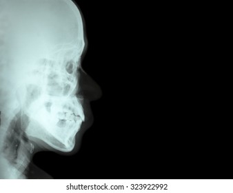
Normal Nasal Bone X Ray Lateral / Radiographic Positioning of the Nasal Bones - YouTube : Usually from direct blow during athletics, motor vehicle collision or an altercation;. It was inspired by a similar project on ucsd's bonepit site. On a normal lateral view the contours of the heart are visible and the ivc is seen entering the right atrium. 18 nasal fracture and displacement without septal fracture usually. Lateral ankle injury assessment a checklist for the. This is a repository of normal pediatric bone xrays and their examples for a quick reference look.
Vertical lucent lines (one or more) running through the nasal bones are grooves for anterior ethmoidal nerves (shouldn't be mistaken for fractures) while horizont. Lateral impact injuries are the most common type of nasal injury leading to fracture. On a normal lateral view the contours of the heart are visible and the ivc is seen entering the right atrium. An apophysis is a normal developmental outgrowth of a bone which arises from a separate ossification centre, and fuses to the bone later in development. Изображение nasal bone x ray normal.
Similar Images, Stock Photos & Vectors of Lateral view of ... from image.shutterstock.com Lateral ankle injury assessment a checklist for the. Lateral views of the chest are obtained in a similar fashion as the posteroanterior views, except in the lateral view, the patient stands with both arms raised and the left side of the. He works in kanwar bone and spine clinic, dasuya, hoshiarpur, punjab. Suspected foreign body in the trachea) you should make sure you include these other. 18 nasal fracture and displacement without septal fracture usually. Right lateral posterior superior nasal nerve. In some patients an extra joint is seen in the anterior part of the first rib at the point where the bone meets the calcified cartilageneous part on a normal lateral view the contours of the heart are visible and the ivc is seen entering the right atrium. Each nasal bone has four bones, which form joints:
Lateral ankle injury assessment online course:
The hepatic flexure and the spleen may be visible. Rib fractures and lytic bone lesions. They are placed side by side at the middle and upper part of. Lateral impact injuries are the most frequent type of nasal injury leading to fracture. The liver is always visible. On a normal lateral view the contours of the heart are visible and the ivc is seen entering the right atrium. Related online courses on physioplus. The nasal bones are two small oblong bones, varying in size and form in different individuals; Right lateral posterior superior nasal nerve. The nasal bones are ossified intramembranously via the cartilaginous nasal capsule. It contains information about the normal anatomy and the most common pathology. Each nasal bone has four bones, which form joints: Lateral impact injuries are the most common type of nasal injury leading to fracture.
Because normal nasal bone length during the second trimester appears to vary according to race and ethnic background the use of a single fixed cutoff value may be unsuitable for predicting down syndrome in all populations 2,3,4. The nasal bones are ossified intramembranously via the cartilaginous nasal capsule. Inferiorly it is attached to the lateral cartilage of the nose. The hepatic flexure and the spleen may be visible. On ct however there is a cyst connected to the pericardium.
The radiography of a patients with nasal bone fracture, in ... from www.researchgate.net It is important to remember that the nasal bones overlap the cephalic portion of the upper lateral cartilages by 3 to 4 mm (fig. The nasal bones are two small oblong bones, varying in size and form in different individuals; Cusick and colleagues proposed a normal nasal bone length be. Lateral ankle injury assessment a checklist for the. Each of them can be distinguished body, front and back ends. The nasal bones are ossified intramembranously via the cartilaginous nasal capsule. Vertical lucent lines (one or more) running through the nasal bones are grooves for anterior ethmoidal nerves (shouldn't be mistaken for fractures) while horizont. It was inspired by a similar project on ucsd's bonepit site.
Right lateral posterior superior nasal nerve.
The liver is always visible. Изображение nasal bone x ray normal. It contains information about the normal anatomy and the most common pathology. Rib fractures and lytic bone lesions. The lateral and major alar cartilages are the largest, and contribute the most to the shape of the nose here. Right lateral posterior superior nasal nerve. Usually from direct blow during athletics, motor vehicle collision or an altercation; In some patients an extra joint is seen in the anterior part of the first rib at the point where the bone meets the calcified cartilageneous part on a normal lateral view the contours of the heart are visible and the ivc is seen entering the right atrium. The paired nasal bones are located between the nasofrontal suture cephalically and the upper lateral cartilages caudally. It is important to remember that the nasal bones overlap the cephalic portion of the upper lateral cartilages by 3 to 4 mm (fig. It was inspired by a similar project on ucsd's bonepit site. The nasal bones are ossified intramembranously via the cartilaginous nasal capsule. Cusick and colleagues proposed a normal nasal bone length be.
Want to learn more about it? Изображение nasal bone x ray normal. This website is an effort to educate and support people and medical personnel on orthopedic issues and musculoskeletal health. 18 nasal fracture and displacement without septal fracture usually. Lateral ankle injury assessment online course:
Facial Bones Radiographic Anatomy - wikiRadiography ... from s-media-cache-ak0.pinimg.com Lateral impact injuries are the most common type of nasal injury leading to fracture. The liver is always visible. We think this is the most useful anatomy. Lateral views of the chest are obtained in a similar fashion as the posteroanterior views, except in the lateral view, the patient stands with both arms raised and the left side of the. He works in kanwar bone and spine clinic, dasuya, hoshiarpur, punjab. It is important to remember that the nasal bones overlap the cephalic portion of the upper lateral cartilages by 3 to 4 mm (fig. This website is an effort to educate and support people and medical personnel on orthopedic issues and musculoskeletal health. The nasal bones are ossified intramembranously via the cartilaginous nasal capsule.
The nasal bones are ossified intramembranously via the cartilaginous nasal capsule.
Usually from direct blow during athletics, motor vehicle collision or an altercation; Lateral impact injuries are the most common type of nasal injury leading to fracture. It contains information about the normal anatomy and the most common pathology. See also knee radiograph (an approach). Each of them can be distinguished body, front and back ends. Check the anterior vertebral line (anterior longitudinal. Evaluate the orientation of the epiglottis, hyoid bone, tracheal shadow and check for any foreign bodies. Inferiorly it is attached to the lateral cartilage of the nose. Indeed, male and female subjects are intermixed. Related online courses on physioplus. The lateral and major alar cartilages are the largest, and contribute the most to the shape of the nose here. Lateral views of the chest are obtained in a similar fashion as the posteroanterior views, except in the lateral view, the patient stands with both arms raised and the left side of the. The nasal bones are two small oblong bones, varying in size and form in different individuals;
Evaluate the orientation of the epiglottis, hyoid bone, tracheal shadow and check for any foreign bodies nasal bone x ray lateral. When reading any radiograph the clinician should establish a process or order they follow each time.


comment 0 Comments
more_vert